|
MRIcro is a freeware image processing program available at: http://www.sph.sc.edu/comd/rorden/mricro.html Download the program mricro.zip and the tutorial, mricro_tutorial.zip. It is easy to install and use for both image conversion and masking. If you are an SPM user, it is worth putting a copy on your Windows, Linux or Solaris machine.Take a look at Chris's excellent tutorials and links to other software online to learn more. Image Viewing and Conversion Providing you have MRIcro installed, here is how you can take a set of MR files and convert them into an ANALYZE/SPM format *.img and *.hdr files. A) Start mricro B) Import-->Convert Dicomm/Genesis/Interfile/Siemens/Picker to Analyze (In the following case we have 17 *.MR files for the 2D/T1 image)
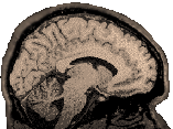
MRIcro will ask you for the first file in the series. Browse for and find the first *.MR file (*1.MR). By default, it will save the *.img and *.hdr file it produces as *1.img and *1.hdr, but it will ask you what you want to name the file and where to put it, so you can tell it at that point if you'd like to make a different choice. Note two things about the program: It does not flip the image upside down (t12spm results in an eyeballs-down axial image, but MRIcro results in an eyeballs-up axial image). It does a left-right flip (i.e., our images are switched from radiological into neurological orientation). You may find your sagittal view is upside down. I found I could fix this by checking "First file is Dorsal" in the panel where I chose the conversion characteristics (see "B" above). If you do this, your image will stay in your original L-R orientation (i.e., our images remain in radiological orientation). If you want your image to match the spm template and you are beginning with images in radiological orientation, then you need to check "flip L-R" in conjunction with "First File is Dorsal" Visit the Image Properties page for more information on image orientation and the effects of various flips and conversions. If you have an image file that you have converted to spm format using phx2spm.m or efl2ana (aka t12spm.m and t3d2spm.m), MRIcro will get an error reading it. It seems to believe these images are 8-bit. You can correct this problem by first trying to open the image, and then when it fails, altering the header to say "16 bit" instead of "8 bit" (the header info appears in the upper left panel). MRIcro will complain that the image has been unloaded from memory when you go to change the header info, but forge ahead. Press the little icon that looks like a floppy disk with "hdr" written across it and overwrite your old header. Now you should have no problem reopening the image. This works with both the structural and functional images. View images with MRIcro by choosing "File-->Open Analyze format hdr and img" and selecting the header or simply drag the image out of the windows explorer menu onto the MRIcro window. You should see an axial image. In the "slice viewer subsection" move the first slider to move through the slices. In the second row of icons in the lower part of this subsection the leftmost brain icon is axial (and selected). To its right are the sagittal, and coronal icons. Click the sagittal and coronal icons and move through the slices. The images may appear dark. If so, hit F5 or choose 'View' and 'Contrast Autobalance'. Check out the "save as" button. This will allow you to convert the image from 16 to 8 bits (for example), or you can tell MRIcro if the default image is something other than axial. |